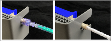
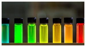
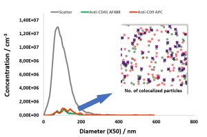
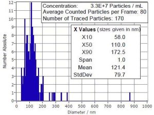
Analysis of 25 nm functionalized QDs using fluorescence-NTA with ZetaView® QUATT system
Application Note Download Abstract While it is uncertain how far the NTA community is able to push the limits of current technology in order to
0x consumables
5x faster switching
10x faster cleaning
12x fluorescence channels
∞x statistics
…and much more!
We measure your products in a measuring range of 0.8 nm to 300 μm, if possible in original concentration
The PARTICLE METRIX Scanning ZetaView® can now be equipped with 3 different Cell Assemblies: “Z NTA” is the renamed classical multi-parameter Cell Assembly including electrophoresis zeta potential. The new innovation is the Cell Assembly for potentially contaminating samples, called “X NTA”, in which all analyses are possible with exception of zeta potential. The same capability is offered with the Standard “NTA” Cell Assembly, which can be used with standard non contaminating samples.
Table 1 lists the performance of all three Cell Assemblies.
For the Z-NTA the classic cell arrangement remains unchanged but has a new name “Z-NTA” to differentiate it from the new Cell Assemblies. It has zeta potential capability as usual.
The new cell units consist of a reinforced quartz cell and its colored holder. The blue holder is designated for the standard (Fig. 1) and the red one for the X-NTA Assembly. Both new Cell Assemblies are designed for rapid routine use like concentration and size analysis experiments. Furthermore, fluorescence detection is possible.
The Cell Assemblies are designed against vibration and drift. Valves at both ends are disposable and ensure leaking free operation.
Other functions of the cell assemblies are listed in Table 2.
The following functions of the ZetaView™ instrument are disabled when using NTA and X-NTA Cell Assemblies: Electrophoresis zeta potential, conductivity and automatic rinsing / dosing via the internal pumps.
The Standard NTA (shown in Fig. 2) and the X-NTA Cell Assembly suit onto the standard ZetaView™ instrument Model PMX110.
Table 3 lists the materials in direct contact with the sample fluid.
Standard NTA Cell Unit on blue holder X-NTA Cell Unit on red holder (Fig. 3).
The new quartz Cell Unit of the Standard NTA and X-NTA assembly is reinforced and therefore very robust. The Cell Unit normally stays in the Assembly, even for cleaning. However, the user may remove the Cell Unit from the Cell Assembly by removing one screw, which is done within seconds (Fig. 5).
Before cleaning, rinse with more than 5 mL of buffer or water, by using a syringe. Afterwards flush with water to remove any sample residues. Remove check valve on the injection side. Dilute cleaning detergent (e.g. 1 mL liquid soap in 50 mL) in water. Wet the pipe cleaner with the cleaning liquid. Insert pipe cleaner directly into the injection port WITHOUT check valve (Fig. 6 right). Fill at least 10 mL of clean water to remove the cleaning solution. The Cell Assembly can now be placed back onto the instrument and locked with a click.
Particle Metrix GmbH is a company producing nanoparticle analyzers to measure size, concentration, zeta potential, fluorescence and pattern sub-populations. All instruments are complementary to each other in size and concentration range. Our design focusses on efficient and secure analysis.
Application Note Download Abstract While it is uncertain how far the NTA community is able to push the limits of current technology in order to
After successfully showing C-NTA of double-stained MSC-EVs in our previous study (https://bit.ly/43GG0Yk) and confirming
the results with image flow cytometry, this note describes C-NTA of multi-stained MSC-EVs using a PMX-430 QUATT NTA
system with four lasers.
In this new application note we describe a quick determination of the physical titer of coronaviruses by fluorescence-based Nanoparticle Tracking Analysis and specific spike antibody recognition.
In this report, we describe the quantification of colocalization ratios on double-stained MSC-derived EVs using fluorochrome conjugated antibodies against the
cell surface antigens CD9 and CD41, which also serve as EV marker proteins.
In addition to using Nano Particle Tracking Analysis (NTA) to measurement the size distribution and concentration of EV samples, both Microfluidic Resistive Pulse Sensing (MRPS) and the Single Particle Interference Reflectance Image Sensor (SP-IRIS) methods have been widely used as an alternative means of characterizing EVs. In this note, we relay two specific cases for using the ZetaView® NTA system to achieve relative “high-throughput” analysis of many EV samples, along estimations of throughput for MRPS & SPIRIS methods for an equivalent number of samples; further, we establish realistic estimates for the high cost of ownership for operating MRPS & SP-IRIS systems as a result of the cost of consumables as well as the substantially greater amount of time spent to run the same number of samples.
Along with generally accepted methods are some “tricks of the trade” such as the addition of Tween® or BSA; however, some of those additions are problematic.
Headquarter
Support
Opt-out complete; your visits to this website will not be recorded by the Web Analytics tool. Note that if you clear your cookies, delete the opt-out cookie, or if you change computers or Web browsers, you will need to perform the opt-out procedure again.
You may choose to prevent this website from aggregating and analyzing the actions you take here. Doing so will protect your privacy, but will also prevent the owner from learning from your actions and creating a better experience for you and other users.
The tracking opt-out feature requires cookies to be enabled.